Foot & Ankle
Foot & Ankle Anatomy
What is the Normal Anatomy of the Foot and Ankle?
The foot and ankle is a complex joint involved in movement and providing stability and balance to the body. The foot and ankle consists of 26 bones, 33 joints, and many muscles, tendons and ligaments.
Bones of the Ankle
The ankle joint connects the leg with the foot, and is composed of three bones: tibia, fibula and talus. The tibia or shin bone and fibula or calf bone are bones of the lower leg which articulate with the talus or ankle bone, enabling up and down movement of the foot.
Three bony bumps present on the ends of the tibia and fibula form parts of the ankle joint:
- The Medial malleolus, formed by the tibia, is found on the inside of the ankle;
- Posterior malleolus, also formed by the tibia, is found at the back of the ankle and the
- Lateral malleolus, formed by the fibula, is found on the outer aspect of the ankle
Bones of the Feet
The foot acts as a single functional unit, but can be divided into three parts: the hindfoot, midfoot and forefoot.
The hindfoot forms the ankle and heel and is made up of the talus bone and calcaneous or heel bone. The heel bone is the largest bone in the foot.
The midfoot connects the hindfoot to the forefoot, and consists of one navicular bone, one cuboid bone, and three cuneiform bones. The navicular bone is found in front of the heel bone, and the cuneiform and cuboid bones are arranged in front of the navicular bone.
These bones are connected to five metatarsal bones of the forefoot, which form the arch of the foot for shock absorption while walking or running. The forefoot is also made up of the toes or digits, formed by phalanges, three in each toe, except the big toe, which has only two phalanges. The big two has two additional tiny round sesamoid bones in the ball of the foot, which help in upward and downward movement of the toe.
Ankle and Foot Joints
There are 33 joints in the ankle and foot. They include the
- Hinge joints in the ankle, which allow flexion (bending) and extension
- Gliding joints found in the hindfoot, which allow gliding movements
- Condyloid joints found in the forefoot and toes, which allow the flexion (bending) and extension, adduction and abduction (sideward movement).
The joints of the foot and ankle provide stability and support the weight of the body, helping you to walk or run, and to adapt to uneven ground.
The joint surface of all bones of the ankle and foot are lined by a thin, tough, flexible, and slippery surface called articular cartilage, which acts as a shock absorber and cushion to reduce friction between the bones. The cartilage is lubricated by synovial fluid, which further enables smooth movement of the bones.
Soft Tissues of the Ankle and Foot
Our feet and ankle bones are held in place and supported by various soft tissues such as cartilage, ligaments, muscles, tendons and bursae.
Cartilage is the flexible, shiny, smooth tissue on the ends of bones that meet to form a joint. Cartilage provides cushioning between the bones allowing smooth movement.
Ligaments are tough rope-like tissue that connect bones to other bones, and holds them in place providing stability to the joints. The Plantar fascia is the largest ligament in the foot, originating from the heel bone to the forefoot, it extends along the bottom surface of the foot and is involved in maintaining the arch of the foot. The plantar fascia ligament stretches and contracts to provide balance and strength to the foot. Lateral ligaments on the outside of the foot and medial ligaments on the inside of the foot provide stability and allow up and down movement of the foot.
The foot is made up of 20 muscles, which help in movement. The main muscles include:
- Anterior tibial muscle: allows up and down movement of the foot
- Posterior tibial muscle: supports the arch
- Peroneal tibial muscle: controls movement on the outside of the ankle
- Extensors: enable the ankle to raise the toes just before stepping forward
- Flexors: stabilize the toes against the floor
Smaller muscles are also present to help the toes lift and curl.
Tendons are soft tissues that connect muscles to bones. The largest and strongest tendon in the foot is the Achilles tendon, present at the back of the lower leg around the heel bone. Other tendons include peroneals and anterior and posterior tibialis.
Bursae
Bursae are small fluid filled sacs that decrease friction between tendons and bone or skin. Bursae contain special cells called synovial cells that secrete a lubricating fluid.
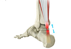
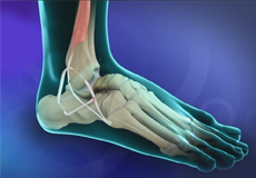
Achilles Rupture
The Achilles tendon is a strong, fibrous cord present behind the ankle that connects the calf muscles to the heel bone. It is used when you walk, run and jump. The Achilles tendon ruptures most often in athletes participating in sports that involve running, pivoting and jumping. Recreational sports that may cause Achilles rupture include tennis, football, basketball and gymnastics.
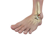
Ankle Fractures
The ankle joint is composed of three bones: the tibia, fibula and talus, which are articulated together. The ends of the fibula and tibia (lower leg bones) form the inner and outer malleolus, which are the bony protrusions of the ankle joint that you can feel and see on either side of the ankle. The joint is protected by a fibrous membrane called a joint capsule, and filled with synovial fluid to enable smooth movement.
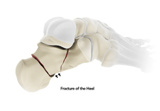
Foot Fractures
Trauma and repeated stress can cause fractures in the foot. Extreme force is required to fracture the bones in the hind foot. The most common type of foot fracture is a stress fracture, which occurs when repeated activities produce small cracks in the bones.
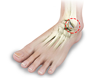
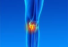
Ankle Arthritis
Arthritis is inflammation resulting from the degeneration of cartilage in the joint causing joint pain, swelling, and stiffness resulting in restricted movements. Arthritis of the foot and ankle joint can occur due to fractures, dislocation, inflammatory disease, or congenital deformity. The foot joints most commonly affected by arthritis are:
Foot Arthritis
Arthritis is inflammation resulting from the degeneration of cartilage in the joint causing joint pain, swelling, and stiffness resulting in restricted movements. Arthritis of the foot and ankle joint can occur due to fractures, dislocation, inflammatory disease, or congenital deformity. The foot joints most commonly affected by arthritis are:
Tendon Tears
The Achilles tendon is a strong, fibrous cord present behind the ankle that connects the calf muscles to the heel bone. It is used when you walk, run and jump. The Achilles tendon ruptures most often in athletes participating in sports that involve running, pivoting and jumping. Recreational sports that may cause Achilles rupture include tennis, football, basketball and gymnastics.
Cartilage Injuries
Coming Soon
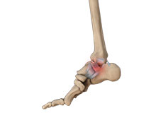
Ankle Sprains
A sprain is the stretching or tearing of ligaments, which connect adjacent bones and provide stability to a joint. An ankle sprain is a common injury that occurs when you suddenly fall or twist the ankle joint or when you land your foot in an awkward position after a jump. Most commonly it occurs when you participate in sports or when you jump or run on a surface that is irregular.
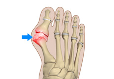
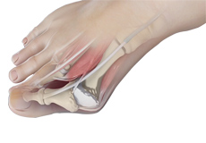
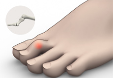
Bunion (Hallux Valgus)
A bunion is a bony protuberance that appears on the outer surface of the big toe when it angles toward the adjacent toe. It is an extra bone and a fluid-filled sac that grows at the base of the big toe.
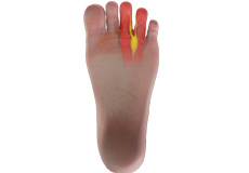
Morton's Neuroma
Morton’s neuroma refers to a nerve injury between the toes, usually the third and fourth toes, which causes pain and thickening of the nerve tissue. Compression or chronic irritation of this interdigital nerve is the main cause of Morton’s Neuroma. Excess pressure is exerted on the nerves due to the narrowing of the gap between the toe bones, causing thickening of the nerve tissue from scar tissue formation. This causes swelling of the nerve and the surrounding tissue.
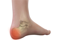
Heel Pain
The heel is made up of the calcaneus bone and supported by a network of muscles, tendons, ligaments and soft tissues, which together support the weight of the body and stress during movement. Heel pain is a common symptom of excessive strain placed on these structures.
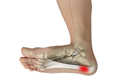
Plantar Fasciitis
Plantar fasciitis refers to inflammation of the plantar fascia, a thick band of tissue that is present at the bottom of the foot. It runs from the heel bone to the toe and forms the arch of your foot. Plantar fasciitis is one of the most common causes of heel pain. It is most often seen in middle-aged men and women, but may also occur in those who are constantly on their feet.
Tibia Fractures
Coming Soon
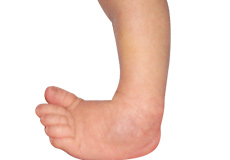
Congenital Deformities
Congenital deformities of the lower limbs are developmental disorders that are present at birth, causing alterations in the shape and appearance of the legs. Several factors such as genetics, teratogenic drugs and chemicals can cause congenital deformities.
Acquired Deformities
Claw toe is a deformity where a toe bends and appears like a bird’s claw. The affected toe is bent upward from the joint at the ball of the foot, and downward at the joints in the middle and tip of the toe to curl under the foot. Hard, thick skin called corns may develop under the ball of the foot or on the top of the affected toe, causing pain while walking.
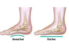
Flat Foot (Pes Planovalgus)
Flatfoot, also known as “fallen arches” or Pes planus, is a deformity in children’s feet in which the arch that runs lengthwise along the sole of the foot has collapsed to the ground or not formed at all. Flatfoot is normal in the first few years of life as the arch of the foot usually develops between the age of 3 and 5 years.
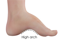
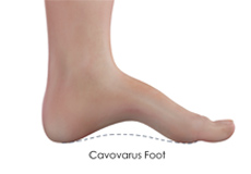
High Arch Foot (Cavovarus Foot)
To support the entire body’s weight on your two feet, the inner middle portion of each foot (midfoot) is raised off the ground to form an arch. A cavovarus foot deformity is characterized by a higher-than-normal arch of the inner midfoot. This results as the two ends of the foot - the heel and toes - abnormally draw towards the inside of the foot, causing the foot to rest on its outer side.
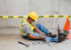
Work Injuries
Injuries at the workplace are very common and may be debilitating. They often occur because of high-risk jobs, lack of appropriate safety devices or lack of training. Common workplace injuries include sprains, fractures, bone dislocations, soft tissue injuries, and injuries requiring limb amputations.
Minimally Invasive Foot/Ankle Surgery
Minimally Invasive Foot Surgery (MIFS) uses the latest advanced technology to treat foot and ankle pain caused by a variety of conditions. Special surgical instruments, devices and advanced imaging techniques are used to visualize and perform the surgery through small incisions. The aim of MIFS is to minimize damage to the muscles and surrounding structures enabling a faster recovery with less pain.
Minimally Invasive Achilles Tendon Repair (Cavovarus Foot)
Tendons are the soft tissues connecting muscle to bone. The Achilles tendon is the longest tendon in the body and is present behind the ankle, joining the calf muscles with the heel bone. Contraction of the calf muscles tightens the Achilles tendon and pulls the heel, enabling the foot and toe movements necessary for walking, running and jumping.
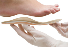
Orthotics
Custom orthotics are specially made devices designed to support and align your feet to address foot-related problems. They are typically created by a healthcare professional such as a podiatrist or orthopedic specialist based on a detailed assessment of your foot structure, gait, and specific needs, often involving a plaster cast, digital scan, or impression of your foot.
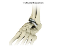
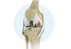
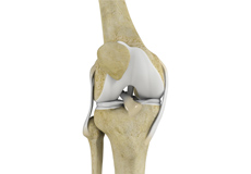
Total Ankle Replacement
The ankle joint connects the leg with the foot and provides free movement to the foot. It is formed by connecting the bones of the lower leg, tibia and fibula, with the talus, or ankle bone.
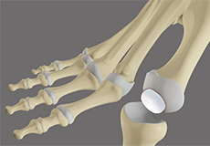
Great Toe Replacement with Synthetic Cartilage
Great toe replacement with synthetic cartilage is a surgical procedure for the treatment of great toe arthritis in which damaged sections of the great toe joint are removed and replaced with an artificial component (synthetic cartilage implant).
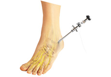
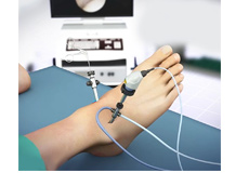
Ankle Arthroscopy
Ankle arthroscopy is a minimally invasive surgical procedure in which an arthroscope, a small, soft, flexible tube with a light and video camera at the end, is inserted into the ankle joint to evaluate and treat a variety of conditions.
Lateral Ankle Ligament Reconstruction
Coming Soon
Bunion Removal
A bunion, also known as hallux valgus, is bony prominence at the base of the big toe, which often results in pain, redness and rubbing in footwear. The first metatarsal bone abnormally angles outward towards the other foot from its joint in the midfoot. A bunion can change the shape of your foot, make it difficult for you to find shoes that fit correctly and worsen the symptoms if left untreated.
Hammer Toe Correction
A hammertoe is a deformity of a lesser toe (second through fifth toes), where the toe is bent upward at the toe’s middle joint, resembling a hammer. The bent portion may rub against a shoe causing pain, irritation and development of corns.