An ankle sprain may seem minor at first, but lingering pain and limited mobility can signal a more complex issue beneath the surface. When symptoms persist for weeks or months, it becomes harder to know whether you’re still healing or dealing with an injury that never fully recovered. Learn when chronic ankle pain warrants an MRI and what conditions it may uncover.
Why Some Ankle Sprains Don’t Fully Heal
Most sprained ankles improve with rest, ice, and protected movement, but not all injuries follow a predictable recovery timeline. Recurrent swelling, stiffness, and instability can indicate underlying damage that routine X-rays cannot detect. Some patients continue to struggle because torn ligaments healed in a stretched position, cartilage was damaged during the original injury, or hidden fractures were never identified.
When ankle pain lingers beyond the expected recovery time, typically six to eight weeks, it may point to deeper structural issues that need evaluation by a foot and ankle specialist.
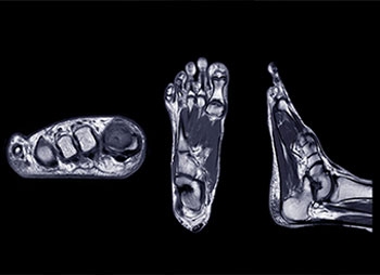
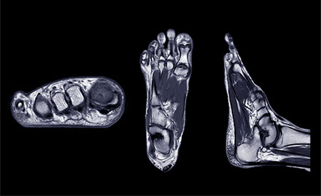
What an MRI Can Reveal About Your Ankle that X-Rays Miss
X-rays are excellent for detecting fractures, but do not show ligaments, cartilage, tendons, or subtle bone bruises. An MRI provides a detailed look at the soft tissues inside the ankle, helping reveal injuries that commonly cause chronic pain. These may include microtears of the lateral ankle ligaments, tendon injuries involving the peroneal tendons, cartilage defects known as osteochondral lesions, or inflammation and scar tissue trapped inside the joint.
Because these conditions often mimic the symptoms of a simple sprain, an MRI becomes a valuable tool when pain persists without a clear explanation.
Signs It May Be Time to Consider an MRI
When ankle pain continues well beyond the normal healing window, certain red flags suggest that an MRI could provide important answers. Persistent swelling that returns with activity, pain along the outer ankle during walking or pivoting, or a sense of instability that makes you cautious on uneven surfaces are possible indicators of hidden damage. Some patients notice clicking or locking inside the joint, while others report deep, nagging discomfort with no improvement despite physical therapy or supportive bracing. These symptoms often point to conditions that require targeted treatment based on MRI findings.
Common Conditions Identified by MRI in Chronic Ankle Pain
An MRI can uncover a number of issues that mimic or complicate a sprain. Ligament injuries may include partial or complete tears of the anterior talofibular or calcaneofibular ligaments, both critical for ankle stability. Tendon problems often involve the peroneal tendons, which can become irritated, torn, or subluxed after injury. Cartilage damage, particularly osteochondral lesions of the talus, can cause sharp pain and swelling that worsens with weight-bearing. In some cases, bone bruises, scar tissue, or subtle ankle fractures appear on MRI even when early imaging seemed normal.
Identifying these problems accurately is essential to developing a treatment plan that restores confidence and prevents long-term weakness or instability.
When to See a Foot and Ankle Specialist
If chronic ankle pain is limiting activity, affecting performance, or simply not improving despite consistent care, it’s worth consulting an orthopedic foot and ankle specialist. An accurate diagnosis helps ensure you receive the right treatment, whether that involves rehabilitation, bracing, minimally invasive procedures, or advanced imaging such as MRI.
For personalized guidance and a clear plan to address persistent ankle pain, book an appointment with Dr. Ho, a foot and ankle specialist who can evaluate your symptoms and help you get back to moving comfortably again.
AUTHOR: Bryant S. Ho, MD is board-certified in orthopedic surgery and is trained in the operative and non-operative management of adolescent and adult foot and ankle disorders. Dr. Ho places a strong emphasis on customizing his care for each patient to ensure successful outcomes. He provides all treatment options, including preventative care, conservative management, and operative intervention.